[:vi]
1. Võng mạc là gì?
Võng mạc là má»™t lá»›p ở táºn trong cùng cá»§a mắt nó hoạt động giống như lá»›p film ở máy ảnh.

Sóng ánh sáng tác động tá»›i mắt rồi há»™i tụ ở trên võng mạc kÃch thÃch lá»›p sợi tháºn kinh hoạt động rồi gá»i thông tin tá»›i não sẽ quyết định chúng ta nhìn thấy gì.
2. Bong võng mạc là gì?
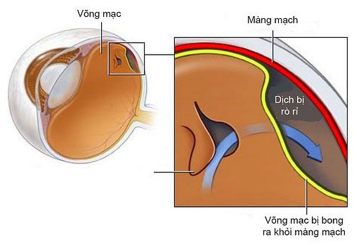
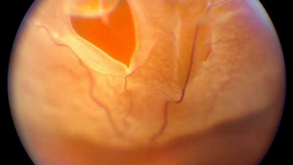
Bong võng mạc có Ä‘iá»u kiện là võng mạc tách lá»›p mô đệm do đó võng mạc mất chức năng.
Có 3 dạng bong võng mạc:
2.1 Bong võng mạc có lỗ rách
Xảy ra thứ phát do sá»± xâm nháºp cá»§a dịch xuống dưới lá»›p võng mạc. Võng mạc tách rá»i so vá»›i lá»›p đệm.

2.2 Bong võng mạc thể tiết dịch
Xảy ra thứ phát do viêm hoặc do vết thương tụ dịch dưới lớp đệm
2.3 Bong võng mạc co kéo
Xảy ra khi sẹo co kéo võng mạc ra khá»i lá»›p mô bên dưới
3. Nguyên nhân của bong võng mạc có lỗ rách
Xảy ra vá»›i tá»· lệ 1/10000 dân số và ở độ tuổi trung niên. Phổ biến hÆ¡n vá»›i những ngưá»i bị cáºn thị và nguy cÆ¡ cao hÆ¡n vá»›i ngưá»i có độ cáºn thị cao hÆ¡n.
Bong võng mạc cÅ©ng có thể xảy ra sau phẫu thuáºt mắt, phổ biến nhất là vá»›i tá»· lệ 0,3%
Chấn thương là một nguyên nhân khác dẫn đến bong võng mạc.
4. Triệu chứng của bong võng mạc
Bong võng mạc thưá»ng bắt đầu bằng sá»± bóc tách thá»§y tinh thể phÃa sau( PVD). Các triệu chứng liên quan đến bóc thá»§y tinh thể ở phÃa sau gồm khởi phát đột ngá»™t và chá»›p sáng. Tuy nhiên, trong má»™t nhóm nhá», tách thá»§y tinh thể ở phÃa sau có thể gây ra rách võng mạc, có thể tiến triển thà nh bong võng mạc.
5. Chẩn đoán bong võng mạc
Một bác sỹ nhãn khoa có thể chẩn đoán được thông qua soi đáy mắt khi đồng tỠgiãn
6. Äiá»u trị có thá»±c sá»± cần thiết?
Bong vá»ng mạc có lá»— rách là má»™t cấp cứu nhãn khoa và phẫu thuáºt là cần thiết để ngăn ngừa mất thị lá»±c vÄ©nh viá»…n.
Mục Ä‘Ãch cá»§a Ä‘iá»u trị là đóng võng mạc bằng laser hoặc liệu pháp áp lạnh (phương pháp đông lạnh), loại bá» dịch dưới võng mạc để là m phẳng nó và há»— trợ võng mạc.

7. Quan Ä‘iểm Ä‘iá»u trị bong võng mạc
7.1 Laser có thể được sá» dụng để Ä‘iá»u trị bong võng mạc và ngăn ngừa sá»± tiến triển cá»§a bong võng mạc. Äiá»u nà y thưá»ng được chá»n nếu bong võng mạc nhá» và cục bá»™, và nếu hoà ng Ä‘iểm không liên quan.
7.2 BÆ¡m hÆ¡i và tạo bóng khà là má»™t kỹ thuáºt trong đó má»™t bong bóng khà được bÆ¡m và o mắt. Bong bóng là má»™t lá»±c chống lại võng mạc tách ra, đẩy nó phẳng và o thà nh trong cá»§a mắt. Äiá»u nà y thÃch hợp trong các trưá»ng hợp được lá»±a chá»n.
7.3 Phẫu thuáºt Ä‘ai độn cá»§ng mạc là phẫu thuáºt sá» dụng Ä‘ai/độn silicon ép cá»§ng mạc từ phÃa ngoà i giúp võng mạc ép và o cá»§ng mạc được dá»… dà ng. PT nà y thưá»ng kèm theo bÆ¡m khà ná»™i nhãn để kết hợp ép võng mạc từ phÃa trong và lạnh đông cá»§ng mạc.
[:en]
1. What is retina?
The retina is the innermost layer of the eye that acts like the film in a camera.

Light waves affect the eye and then focus on the retina to stimulate the nerve fiber layer to work and then send information to the brain that will determine what we see.
2. What is retinal detachment?
Conditional retinal detachment is when the retina separates from the stroma and thus loses its function.
There are 3 types of retinal detachment:
2.1 Retinal detachment with laceration
Occurs secondary to infiltration of fluid below the retinal layer. Retina separates from the stroma.

2.2 exudative retinal detachment
Occurs secondary to inflammation or due to wound accumulation of fluid under the buffer
2.3 Traction retinal detachment
Occurs when the scar pulls the retina away from the underlying tissue
3. Causes of retinal detachment with holes
Occurs with the rate of 1/10000 of the population and in middle age. More common with people with myopia and higher risk with people with higher levels of myopia.
Retinal detachment can also occur after eye surgery, most commonly with an incidence of 0.3%
Trauma is another cause for retinal detachment.
4. Symptoms of retinal detachment
Retinal detachment usually begins with posterior vitreous detachment (PVD). Symptoms associated with posterior vitreous detachment include sudden onset and flash of light. However, in a small group, posterior vitreous detachment can cause retinal tear, which can progress to retinal detachment.
5. Diagnosing retinal detachment
An ophthalmologist can diagnose by ophthalmoscopy when the pupils are dilated
6. Is treatment really necessary?
A blepharoplasty is an ophthalmic emergency and surgery is necessary to prevent permanent vision loss.
The goal of treatment is to close the retina with laser or cryotherapy (freezing method), removing fluid under the retina to flatten it and support the retina.

7. Opinion on treatment of retinal detachment
7.1 Laser can be used to treat retinal detachment and prevent progression of retinal detachment. This is usually chosen if the retinal detachment is small and local, and if the macula is not involved.
7.2 Inflating and ballooning is a technique in which an air bubble is injected into the eye. The bubble is a force against the detached retina, pushing it flat against the inner wall of the eye. This is appropriate in selected cases.
7.3 Scleroplasty is a surgery that uses a silicone belt/filler to squeeze the sclera from the outside to help the retina press against the sclera easily. This surgery is usually accompanied by intraocular gas injection to combine retinal compression from the inside and scleral freezing.
[:]